The term onychomycosis (toenail and fingernail fungus) describes fungal nail infections caused by non-dermatological skin fungi, molds, or yeasts. There are four clinically distinct forms of onychomycosis. Diagnosis is based on examination by CON, microscopy and histology. Typically, treatment includes systemic and local therapy, sometimes requiring surgical excision.

Factors that contribute to nail fungus
- Increased sweating (hyperhidrosis).
- Vascular insufficiency. Violation of the structure and tone of the veins, especially the veins of the lower limbs (typical onychomycosis of the toenails).
- Year old. The incidence of the disease in humans increases with age. In 15-20% of the population, pathology occurs at the age of 40-60.
- Diseases of internal organs. Disruption of the nervous system, endocrine system (most commonly onychomycosis occurring in people with diabetes) or immune system (immunosuppression, especially with HIV infection).
- Large hoof masses, consisting of a thick nail plate and the material underneath it, can cause discomfort when wearing shoes.
- Injury. Continuous injury to the nail or trauma and lack of appropriate treatment.
Disease incidence
Nail fungus– the most common nail disease, the cause of 50% of cases of nail dystrophy (destruction of the nail plate). It affects up to 14% of the population and both its incidence in the elderly and its overall incidence are increasing. The incidence of onychomycosis in children and adolescents is also increasing, with onychomycosis accounting for 20% of fungal skin infections in children.
The increase in incidence may be related to wearing tight shoes, increased numbers of people taking immunosuppressive therapy, and increased use of public locker rooms.
Nail disease often begins with athlete's foot before spreading to the nail bed, where it is difficult to remove. This area serves as a reservoir for local recurrences or spread of the disease to other areas. Up to 40% of patients with toe nail fungus have associated skin infections, most commonly athlete's foot (about 30%).
Causative agent of nail fungus
In most cases, onychomycosis is caused by dermatophytes, with T. rubrum and T. interdigitale being the infecting agents in 90% of cases. T. tonsurans and E. floccosum have also been reported as pathogens.
Yeasts and non-dermatophyte mold organisms such as Acremonium, Aspergillus, Fusarium, Scopulariopsis brevicaulis and Scytalidium are the source of toenail fungus in about 10% of cases. It is interesting to note that Candida species are the causative agents in 30% of cases of finger onychomycosis, while non-cutaneous molds are not found in affected nails.
Pathogenesis
Dermatophytes have a variety of enzymes, which act as virulence factors, ensuring the adhesion of pathogens to the nail. The first stage of infection is adhesion to keratin. Due to further breakdown of keratin and release of downstream mediators, an inflammatory response develops.

The pathogenesis stages of fungal infections are as follows.
Adhesion
The fungus overcomes several lines of host defense before the hyphae begin to exist in the keratinized tissues. The first is the successful adhesion of Arthroconidia to the surface of keratinized tissues. The host's initial nonspecific lines of defense include fatty acids in sebum, as well as colonization by competing bacteria.
Several recent studies have examined the molecular mechanisms involved in the adhesion of arthroconidia to keratinized surfaces. Dermatocytes have been shown to selectively utilize their proteolytic reserves during adhesion and invasion. Some time after attachment takes place, the spore germinates and moves on to the next stage - invasion.
Invasion
Trauma and rubbing are favorable environments for fungal invasion. The colonization of fungal germination factors ends with the release of different types of proteases and lipases, in general different products that serve as nutrients for the fungus.
Owner's reaction
Fungi face multiple defenses in the host, such as inflammatory mediators, fatty acids, and cellular immunity. The first and most important barrier is the keratinocytes, which are encountered when fungal elements enter. Role of keratinocytes: proliferation (increased keratin shedding), secretion of antibacterial peptides, anti-inflammatory cytokines. As the fungus penetrates deeper, more and more new non-specific mechanisms are activated for protection.
The severity of the host's inflammatory response depends on the immune status, as well as the natural habitat of the dermatophytes participating in the invasion. The next level of defense is the delayed-type hypersensitivity reaction, caused by cell-mediated immunity.
The inflammatory response associated with this hypersensitivity is associated with clinical destruction, while defects in cell-mediated immunity can lead to chronic and recurrent fungal infections.
Although epidemiological observations suggest a genetic predisposition to fungal infections, none have been demonstrated at the molecular level.
Clinical images and symptoms of toenail and fingernail damage
There are four characteristic clinical forms of infection. These forms may be separate or include several clinical forms.
Distal sublingual onychomycosis
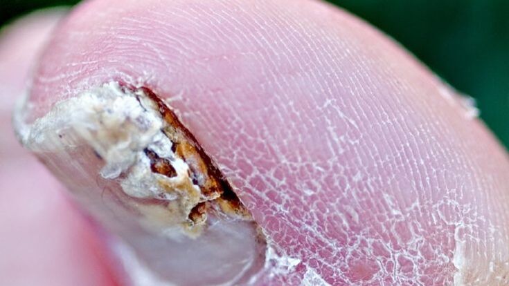
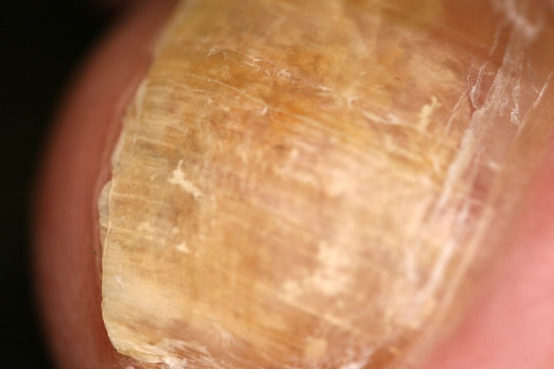
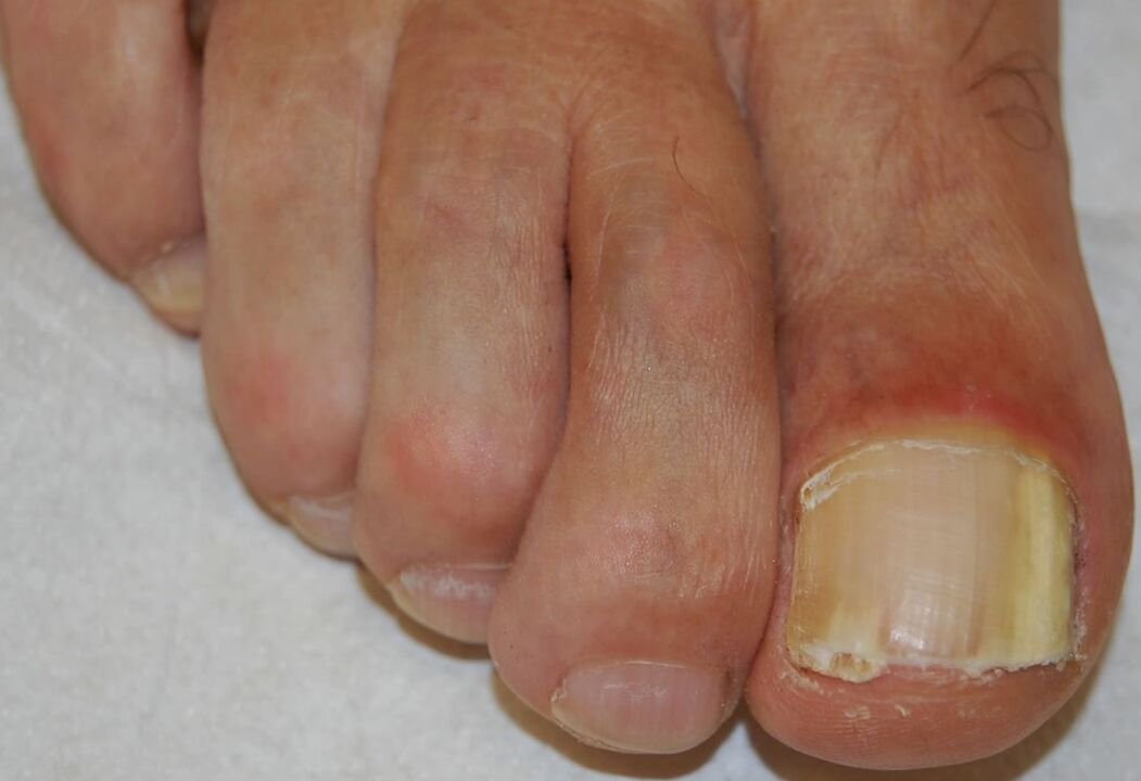
This is the most common form of nail fungus and can be caused by any of the pathogens listed above. It begins with pathogen penetration into the stratum corneum of the hypoungual and distal part of the nail, resulting in a white or yellow-brown opacification of the distal tip of the nail. The infection then spreads gradually up the lower part of the nail to the ventral surface of the nail plate.

Excess proliferation or impaired differentiation of the nail bed in response to infection causes subungual hyperkeratosis, while progressive invasion of the nail plate results in increased nail dystrophy.
Onychomycosis under the proximal nail
It occurs due to infection of the proximal nail fold, primarily by the organisms T. rubrum and T. megninii. Clinic: the proximal part of the nail is white or beige. This opacification gradually increases and affects the entire nail, eventually leading to leukemia, nail necrosis, and/or total nail destruction.
Patients with proximal subungual onychomycosis should be tested for HIV infection, as this form is considered a sign of this disease.
White surface onychomycosis
It occurs by direct invasion of the back of the nail and appears as clear, white or dull yellow spots on the surface of the toenail. The most common pathogens are T. interdigitale and T. mentargophytes, although non-dermatophyte molds such as Aspergillus, Fusarium and Scopulariopsis are also known to be such pathogens. Candida species can penetrate the subungual area of the epithelium and eventually infect the nail along the entire thickness of the nail plate.
Nail fungus caused by Candida
Nail plate damage caused by Candida albicans is observed only in chronic mucosal candidiasis (a rare disease). Usually all nails are affected. The nail plate thickens and acquires various shades of yellow-brown color.
Diagnosis of nail fungus
Although onychomycosis accounts for 50% of nail dystrophy cases, laboratory results confirming the diagnosis are recommended before starting toxic systemic antifungal medications.
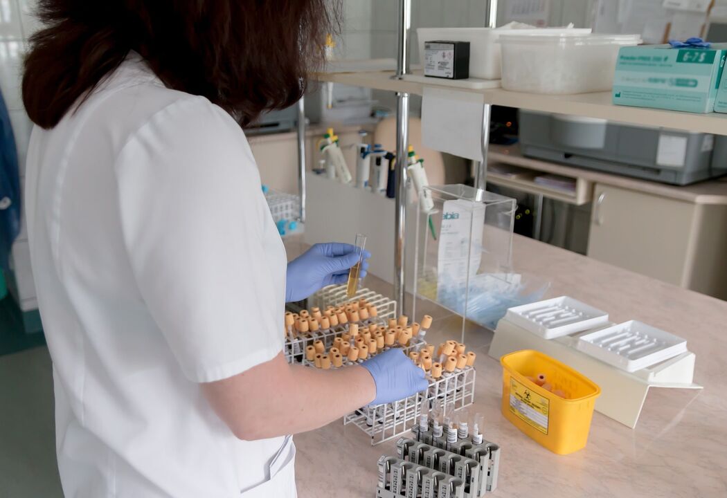
Study of the subungual mass with KOH, material culture analysis of the nail plate and subungual mass on Sabouraud dextrose agar (with and without antibacterial additives) and nail staining by the PAS method are the methodsgives the most information.
Study with CHILDREN
This is a standard test for suspected onychomycosis. However, it is often negative even when there is a high degree of clinical suspicion, and culture analysis of nail material found for fungal hyphae during studies with CON is often negative.
The most reliable way to minimize false negative results due to sampling error is to increase the sample size and resample.
Cultural analysis
This laboratory test identifies the type of fungus and determines the presence of dermatophytes (organisms that respond to antifungal medications).

To distinguish pathogens from contaminants, the following recommendations are given:
- if dermatophyte is isolated in culture it is considered a pathogen;
- Non-tanning mold or yeast organisms isolated in culture are relevant only if hyphae, spores or yeast cells are observed under the microscope and active growth is observed. Recurrence of nondermatophyte mold pathogens without isolation.
Cultural analysis, PAS - the method of nail dyeing is the most sensitive and does not require waiting for the results for several weeks.
Check pathology
During histopathological examination, fungal hyphae are located between the layers of the nail plate, parallel to the surface. In the epidermis, focal spongiosis and parakeratosis can be observed, as well as an inflammatory reaction.
In superficial white onychomycosis, microorganisms are found on the posterior surface of the nail, displaying their unique "perforated organ" pattern and modified hyphal components called "biting leaves". With candidal onychomycosis, pseudofibrous invasion is observed. Histological examination of onychomycosis occurs using special dyes.
Differential diagnosis of nail fungus
| Likely | Sometimes it can happen | Rarely found |
|---|---|---|
|
|
melanoma |
Methods of treating nail fungus
Treatment of onychomycosis depends on the severity of the nail lesion, the presence of associated onychomycosis, and the effectiveness and potential side effects of the treatment regimen. If nail involvement is minimal, topical therapy is a reasonable decision. When combined with dermatosis of the feet, especially against the background of diabetes, it is imperative to prescribe treatment.
Topical antifungals
In patients with involvement distal to the nail or contraindications to systemic therapy, local treatment is recommended. However, we must remember that topical treatment with antifungal drugs alone is not effective enough.
A varnish from the oxypyridone group that is becoming increasingly popular, applied daily for 49 weeks, achieves a cure for mycosis in about 40% of patients and clears the nails (clinical cure) in5% of cases of mild or moderate onychomycosis are caused by skin fungi. .
Although much less effective than systemic antifungals, topical use avoids the risk of drug interactions.
Another drug, specially developed in the form of nail polish, is used 2 times a week. It is representative of a new group of antifungal drugs, morpholine derivatives, active against yeasts, dermatophytes and molds that cause onychomycosis.
This product may have a higher cure rate for fungal infections than previous varnishes; however, controlled studies are needed to determine statistically significant differences.
Antifungal drugs taken orally
Systemic antifungals are required in cases of onychomycosis involving the underlying area or if a shorter course of treatment is desired or a better chance of recovery and cure is desired. When choosing an antifungal medication, one must first consider the cause of the disease, potential side effects, and the risk of drug interactions in each patient.
A drug of the allylamine group, which has a fungistatic and fungicidal effect on dermatophytes, Aspergillus, is less effective against Scopulariopsis. This product is not recommended for candida onychomycosis as it shows variable effectiveness against Candida species.
The standard 6-week dose is effective for most toenail injections, while for toenail injections a minimum of 12 weeks is required. Most side effects are related to digestive system problems, including diarrhea, nausea, taste changes, and increased liver enzymes.
Data indicate that a 3-month continuous dosing regimen is currently the most effective systemic treatment for toenail fungus. The clinical cure rate in various studies was approximately 50%, although the cure rate was higher in patients over 65 years of age.
A drug of the azole group that has a fungicidal effect against dermatophytes, as well as non-dermatophyte mold and yeast organisms. Safe and effective regimens include daily pulse dosing for one week each month or daily continuous dosing, both of which require two months or two treatment cycles for nails and at least three months or threetreatment for toenail damage.
In children, the drug is dosed individually depending on weight. Although the drug has a broader spectrum of effects than its predecessor, studies have shown significantly lower cure rates and higher relapse rates.
Elevated liver enzyme levels occurred in less than 0. 5% of patients during treatment and returned to normal within 12 weeks after stopping treatment.
The drug has a fungistatic effect on dermatological cells, some molds that do not cause skin diseases, and Candida species. This medication is usually taken once a week for 3 to 12 months.
There are no clear criteria for laboratory monitoring of patients taking the above drugs. It is reasonable to perform complete blood count and liver function tests before treatment and 6 weeks after starting treatment.
A drug in the Grisan class is no longer considered standard therapy for onychomycosis due to long treatment times, potential side effects, drug-drug interactions, and relatively low cure rates.
Combination treatment regimens may produce higher clearance rates than systemic or topical treatment alone. The use of allylamine combined with application of morpholine varnish resulted in clinical cure and negative fungal testing in approximately 60% of patients, compared with 45% of patients receiving systemic allylamine antifungals alone. However, another study showed no additional benefit when combining systemic allylamine with oxypyridone drug solution.
Other medications
The fungicidal activity demonstrated in vitro for thymol, camphor, menthol, and eucalyptus citriodora oils suggests potential for additional therapeutic strategies in the treatment of onychomycosis. Thymol alcohol solution can be used in drops on the nail plate and on nail beds. The use of topical nail preparations containing thymol has resulted in cure in some isolated cases.
Surgery
The final treatment option for refractory cases includes urea nail excision. To remove more debris from the affected nail, special pliers are used.
Many doctors believe that the main and first method of treating nail fungus is mechanical nail removal. Usually, surgical removal of the affected nail is recommended and, less often, removal using a keratolytic patch.
Traditional methods in the fight against nail fungus
Although there are many different folk recipes for eliminating nail fungus, dermatologists do not recommend choosing this treatment option and starting with "home diagnosis". It would be wiser to start treatment with local drugs that have undergone clinical trials and have been proven to be effective.
Course and prognosis
Signs of poor prognosis include pain caused by thickening of the nail plate, secondary bacterial infection, and diabetes. The most beneficial way to reduce the likelihood of recurrence is to combine treatments. Treating nail fungus is a long road that does not always lead to complete recovery. However, do not forget that the effectiveness of systemic therapy is up to 80%.
Prevent
Prevention includessome events, thanks to which you can significantly reduce the incidence of fungal nail infections and reduce the possibility of recurrence.
- Disinfect personal and public items.
- Disinfect shoes systematically.
- Treat feet, hands, folds (in favorable conditions - preferred location) with topical antifungals as recommended by a dermatologist.
- If the diagnosis of onychomycosis is confirmed, follow-up visits to the doctor are required every 6 weeks and after completion of systemic therapy.
- If possible, every time you visit your doctor, you should have your nail plates cleaned.
Conclusion
Onychomycosis (fingernail and toenail fungus) is an infection caused by many different types of fungi. This disease affects the nail plates of the fingers or toes. When making a diagnosis, examine all skin and nails, and rule out other diseases that resemble onychomycosis. If there is any doubt about the diagnosis, it should be confirmed by culture (preferably) or by histological examination of nail clippings followed by staining.
Therapy includes surgical excision, topical medications, and general medications. Treatment of nail fungus is a long process, which can last several years, so you should not expect recovery "from just one pill". If you suspect nail fungus, consult a specialist to confirm the diagnosis and prescribe an individualized treatment plan.


